


.jpg?width=1024&height=576&name=vRad-High-Quality-Patient-Care-1024x576%20(1).jpg)


%20(2).jpg?width=1008&height=755&name=Copy%20of%20Mega%20Nav%20Images%202025%20(1008%20x%20755%20px)%20(2).jpg)


 vRad Marketing
1 min read
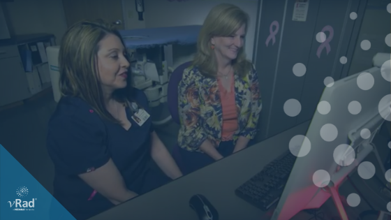
10 Steps to a Stress-Free Mammography Inspection
Whoever said the only things certain in life are death and taxes never faced an MQSA inspection. Federally mandated in the early ’90s...

.png)
1 min read
Whoever said the only things certain in life are death and taxes never faced an MQSA inspection. Federally mandated in the early ’90s...

1 min read
In this blog series Part 3, I will be sharing how newer screening tools now exist including breast MRI.
1 min read
On September 28, 2016 vRad announced that it implemented its first breast imaging Live Video Diagnostics solution site at the Center for Women’s...
vRad (Virtual Radiologic) is a national radiology practice combining clinical excellence with cutting-edge technology development. Each year, we bring exceptional radiology care to millions of patients and empower healthcare providers with technology-driven solutions.
Non-Clinical Inquiries (Total Free):
800.737.0610
Outside U.S.:
011.1.952.595.1111
3600 Minnesota Drive, Suite 800
Edina, MN 55435