


.jpg?width=1024&height=576&name=vRad-High-Quality-Patient-Care-1024x576%20(1).jpg)


%20(2).jpg?width=1008&height=755&name=Copy%20of%20Mega%20Nav%20Images%202025%20(1008%20x%20755%20px)%20(2).jpg)


 J.P. Dym, MD
1 min read
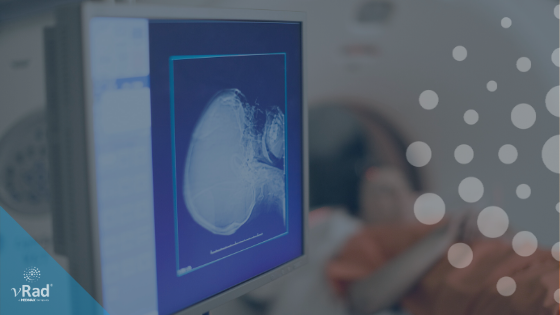
Stroke Awareness Month and vRad’s Impact
May is both Stoke and Trauma awareness month. In recognition of this, we will be publishing a series of 3 posts covering: Stroke information and our...

1 min read
May is both Stoke and Trauma awareness month. In recognition of this, we will be publishing a series of 3 posts covering: Stroke information and our...

1 min read
Today is our third and final post in deference to Stroke and Trauma Awareness Month:

1 min read
As I mentioned in my last post, May is both Trauma and Stroke Awareness month. vRad serves hundreds of trauma and stroke centers across the U.S. The...
vRad (Virtual Radiologic) is a national radiology practice combining clinical excellence with cutting-edge technology development. Each year, we bring exceptional radiology care to millions of patients and empower healthcare providers with technology-driven solutions.
Non-Clinical Inquiries (Total Free):
800.737.0610
Outside U.S.:
011.1.952.595.1111
3600 Minnesota Drive, Suite 800
Edina, MN 55435