


.jpg?width=1024&height=576&name=vRad-High-Quality-Patient-Care-1024x576%20(1).jpg)


%20(2).jpg?width=1008&height=755&name=Copy%20of%20Mega%20Nav%20Images%202025%20(1008%20x%20755%20px)%20(2).jpg)


 Ricardo C. Cury, MD, FACR
1 min read
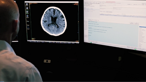
How AI Is Accelerating Care Delivery in Critical Cases
The vRad Imaging Platform employs multiple AI models in processing over 20,000 radiology studies each day. Silently prioritizing critical cases is...

1 min read
The vRad Imaging Platform employs multiple AI models in processing over 20,000 radiology studies each day. Silently prioritizing critical cases is...
.png)
1 min read
“Only a few more months and life will be good!” It’s the rallying cry of radiology residents and fellows everywhere. The idea that if we can just...
.png)
1 min read
At some point in your career, if you are lucky, you find somebody approachable who’s blazed trails and navigated difficulties in ways you can model....
vRad (Virtual Radiologic) is a national radiology practice combining clinical excellence with cutting-edge technology development. Each year, we bring exceptional radiology care to millions of patients and empower healthcare providers with technology-driven solutions.
Non-Clinical Inquiries (Total Free):
800.737.0610
Outside U.S.:
011.1.952.595.1111
3600 Minnesota Drive, Suite 800
Edina, MN 55435