.jpg?width=1024&height=576&name=vRad-High-Quality-Patient-Care-1024x576%20(1).jpg)


%20(2).jpg?width=1008&height=755&name=Copy%20of%20Mega%20Nav%20Images%202025%20(1008%20x%20755%20px)%20(2).jpg)


 Juan Batlle, MD

1 min read
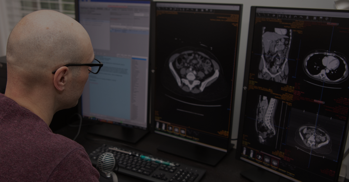
12 Radiologist Safety Nets to Reduce Stress, Improve Care, and Make Radiology Enjoyable Again
This is the first in a series of blog posts we’re calling “@vRad” where radiologists can get a sneak peek behind the curtain to see what it’s like to...